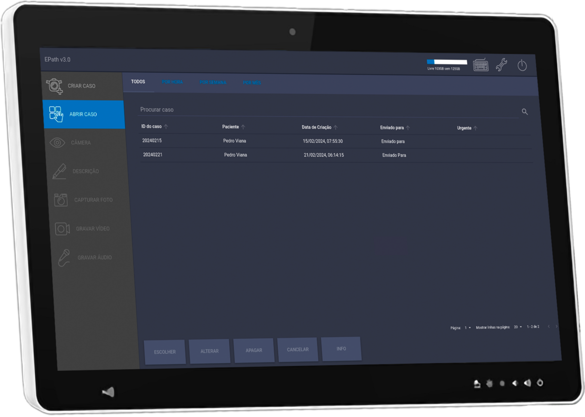
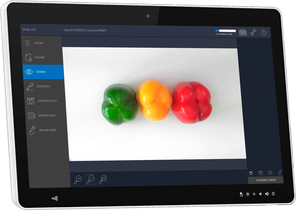
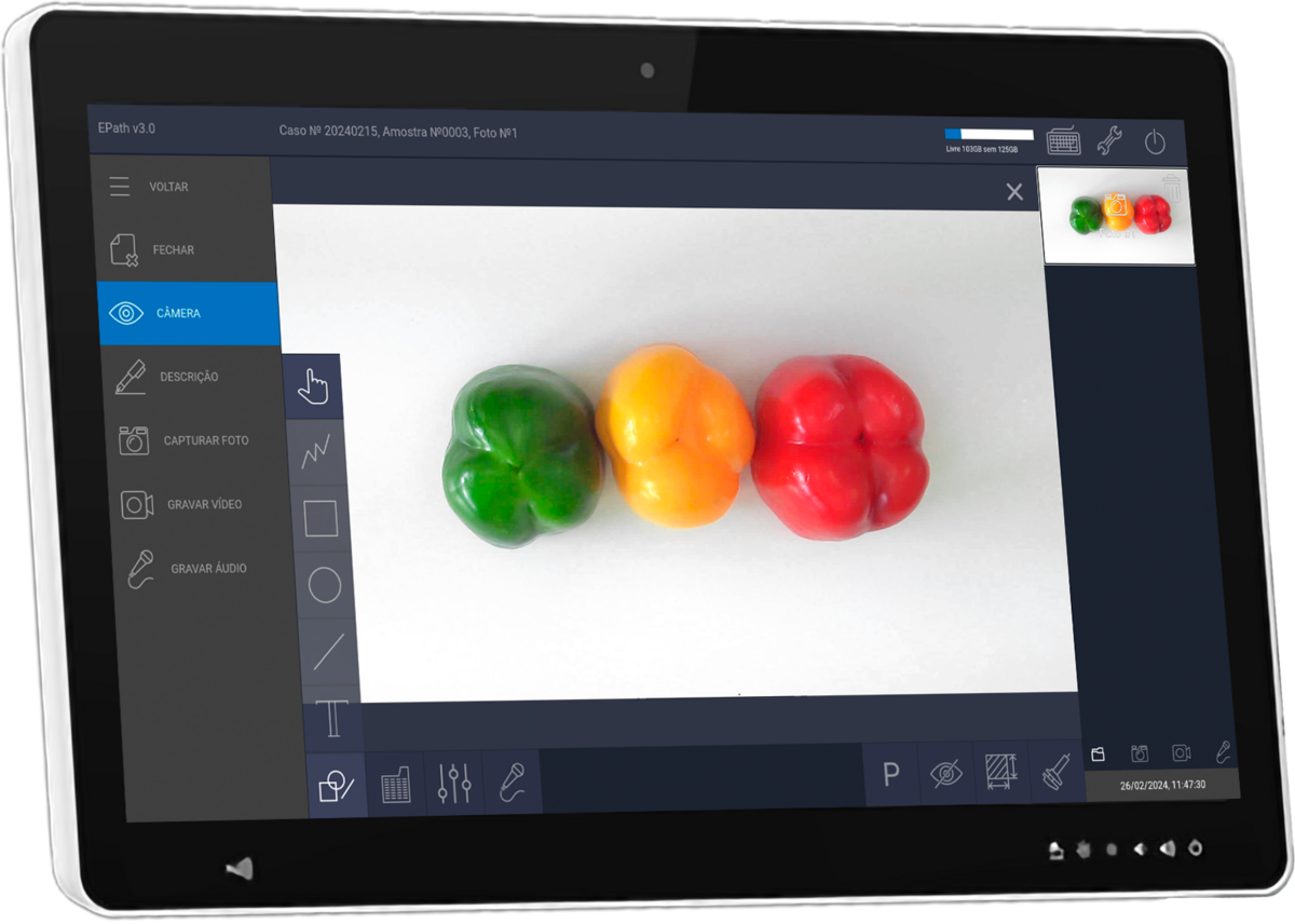
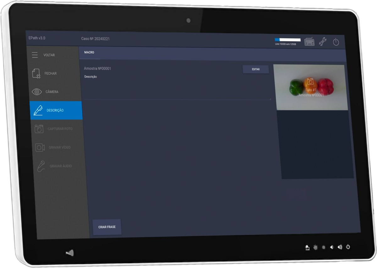
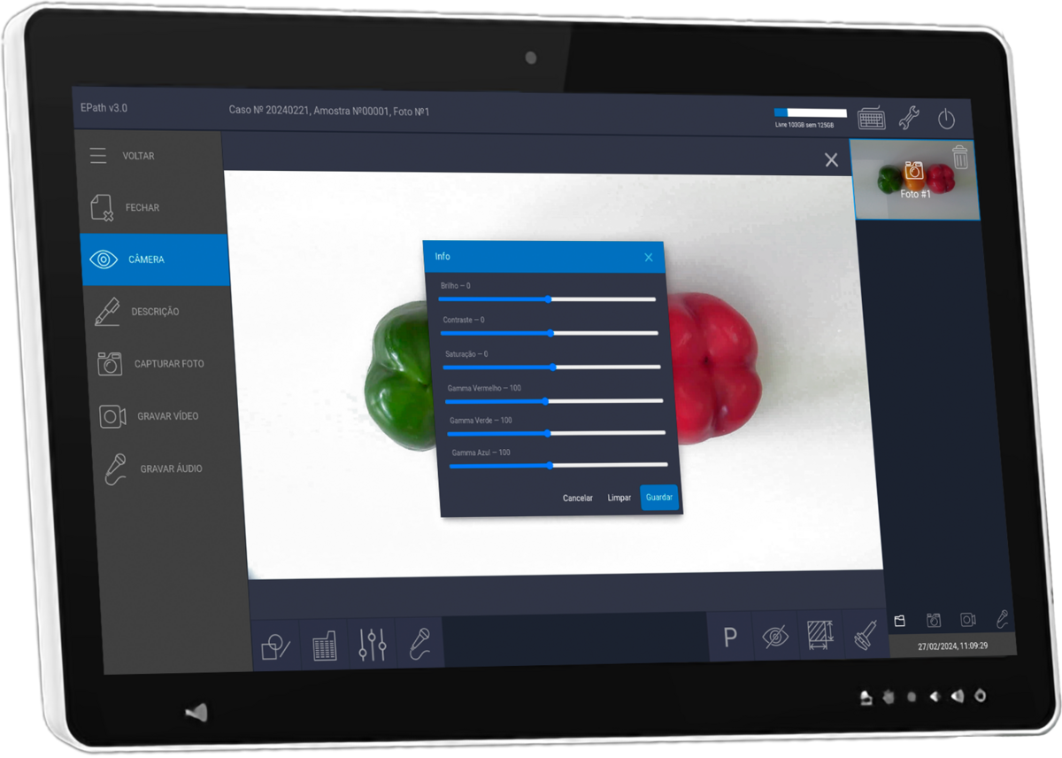
The macroimaging solution EPATH1 allows pathologists to take photos, record videos, and audio during a macroscopic examination procedure.
EPATH can be integrated with DICOM and LIS systems.
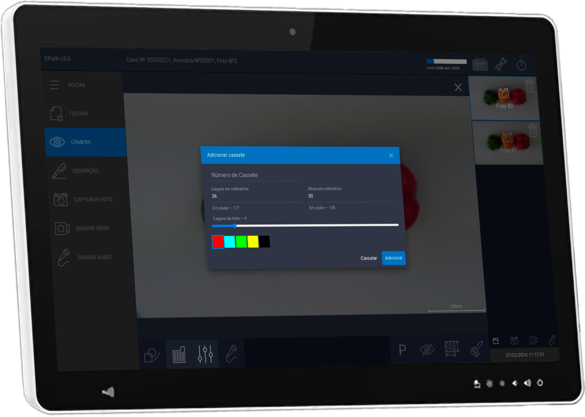
It fully supports pathologists during the macroscopic examination phase of surgical biopsy and autopsy material. The system includes a patented photo and video recording module with automatic laser calibration function, enabling pathologists to capture photos and videos for more accurate morphometric analyses.
High quality photo and video capture.
System control through the use of pedals.
High-quality photo and video capture.
System control through the use of pedals.
Manual white balance settings for different lighting conditions in the laboratory.
Installation on any gross station.
Tools for morphometric analysis: graphical objects in images, such as morphometric figures, linear dimensions, object areas, perimeters, text annotations, histological cassettes
Remote access from any pathologist workstation.
Real-time video streaming of the macroscopic process to remote workstations for real-time control of capture and description, supporting, for example, educational processes.
Automatic laser rangefinder calibration to ensure measurement accuracy for morphometric analysis.
Pre-configured description templates.
Integration with:
LIS (importation of patient case data by reference barcode, ability to open a case in the EPATH interface from the LIS).
Local computer networks of medical organizations to support data export.
DICOM.
1. Do not use the device as a measurement equipment. All data obtained from the process of morphometry might be used for reference only and couldn’t be an only reason to make a diagnosis.